How Do U Know If Retinal Tear Repair Didn't Work
The retina can be likened to moving picture in a camera. It is the light sensitive construction that lines the back of the eye. The part directly in the back of the centre is the macular region. Because of the structure of the macula and the cells that are there, the macula supplies abrupt vision and also provides almost of the color information existence sent back to our brains.
The rest of the retina supplies a lower resolution image that gives u.s.a. the wide field of view we ordinarily have. This side vision is very important in functioning in the modernistic globe. The retina is not connected to the back of the centre in a firm mode. Under some circumstances the retina can pull away from the back of the eye. Since the retina gets much of its oxygen and nutrition from the tissue in the back of the heart, this can lead to significant harm to vision.
Types of Retinal Detachments
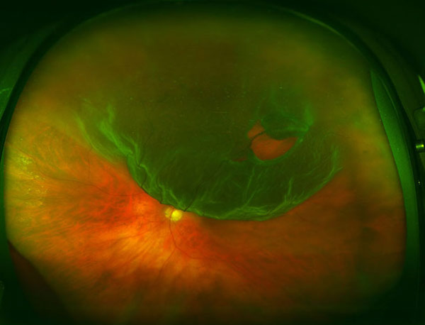
There are many dissimilar ways the retina can detach from the back of the centre. One manner is that it can be pulled by strength. There is no hole or tear in the retina, just animal force. A second way is the retina can tear and fluid from the middle part of the middle can go under the retina; this method combines pulling with fluid flows to cause the retina to split up from the back of the eye. The tertiary important style is there can be an excessive amount of fluid made under the retina by disease and the rising tide of fluid floats the retina away from the back of the eye.
Retinal Disengagement Caused by Pulling: Traction Retinal Detachment
Some diseases, such equally diabetes, cause blood vessels and cells like to those found in scar tissue to grow into the center parts of the eye from the retina. At first these stalks look like treed or fans of seaweed. Eventually they contain more than and more scar. Likewise since they comprise blood vessels these fronds tin also bleed. Bleeding commonly leads to more than scar tissue. Contraction of the scar tissue can pull the retina away from the back of the center. Many different weather can lead to the same endpoint. In many patients the simple passage of time causes the vitreous to pull on the retina, at least a lilliputian. In some patients this pulling tin can be extreme and lead to disengagement of the macula.
Retinal Detachment Acquired past a Retinal Defect: Rhegmatogenous Retinal Detachment
Excessive pulling, particularly if concentrated to a small area, tin can cause the retina to tear. Fluid from the vitreous crenel can come in through the pigsty and accumulate under the retina. This type of retinal detachment has an excessively complicated name, a rhegmatogenous retinal detachment. Retinal tears leading to detachment is the about common form of retinal detachment leading to eye surgery. The link between retinal tears and detachments didn't happen that long agone in medical history. There was no way to look into the center until nearly 150 years ago. It took many years to develop methods that allowed examination of the entire retina. Once that happened people realized the retina could detach, and they saw the retina had tears, but thought the tears occurred because the retina was detached, not the other way effectually.
Excessive Leakage nether the Retina: Exudative Retinal Detachment
A complicated internet of claret vessels exists under the retina in a structure called the choroid. The retina has the highest need for oxygen in the body per gram of tissue. If the retina had enough blood vessels to supply this requirement nosotros would have a difficult time seeing because of all the blood vessels. Instead the centre has a dainty blueprint characteristic. A dense network of blood vessels delivers oxygen to the retina from below. This layer is called the choroid, and the choroid has the highest claret flow per gram of tissue in the torso. Problems can arise if the vessels in the choroid get excessively leaky. If you accept ever injured your elbow or human knee yous know the joint can become swollen. The swelling is from fluid leaking from blood vessels. Imagine the choroid – if that becomes inflamed there are many vessels that could leak. Fluid produced in the choroid will make that layer swollen, simply the fluid oozes up nether the retina and accumulates there. In exudative detachments the retina is floats off of the dorsum of the eye.
Symptoms
 The most common symptoms include:
i. Decreased visual vigil
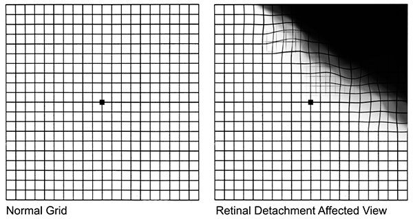
2. Dark regions in the field of vision
three. Distorted vision
Diagnostic Testing
 The principle way doctors at VRM diagnose a retinal detachment is through straight examination of the centre. This tin can assistance identify which of the iii types of causes for retinal disengagement is the problem. Boosted testing is required to evaluate this initial hypothesis. Ultrasound is used to evaluate the position of the retina and to look at deeper structures in the eye.
Optical coherence tomography provides cantankerous-exclusive data about the retina and choroid. Fluorescein angiography is very helpful in grading the amount of leakage in exudative detachments. As in other fields of medicine diagnostic testing helps establish the diagnosis, the mechanism of disease, serves to certificate affliction severity, and helps in monitoring disease progression.
Treatment
Each form of retinal detachment has a different crusade, and to repair the detachment the root causes need to exist addressed.
Traction Retinal Disengagement
Once the source and extent of the traction is visualized, plans can be made to repair the same. In near cases of traction retinal detachment a vitrectomy is performed. This operation removes the vitreous, the jelly-like substance in the heart that typically provides the platform for the scar tissue to grow. In the procedure the tissue providing traction on the retina is likewise removed. Once the traction is removed there is no longer a reason for fluid to accumulate under the retina. However there is already fluid under the retina, since the retina is detached. To get the fluid out from under the retina a minor hole is made in the retina and the fluid is sucked out. The minor hole is covered with laser to weld the retina to the back of the heart. The eye typically is filled with gas to concord the retina in place until the laser weld strengthens.
Rhegmatogenous Detachments: Fixing Detachments with a Retinal Tear
A retinal tear leading to a detachment requires closing the retinal pigsty for the detachment to be repaired. When retinal doctors talk near endmost a hole, what they mean is that the flow of fluid through the retinal tear to the space under the retina must exist blocked.
In that location are iii chief ways to exercise this.
VITRECTOMY
The vitreous and any pulling by the vitreous is removed from the eye. The fluid under the retina is sucked out of the eye and laser welds are put around the tear to seal the tear so no new fluid tin come nether the retina. The centre is usually filled with gas to hold the retina in place until the weld south become stiff. Sometimes a thick fluid, silicone oil, is used instead of gas. The advantage of vitrectomy is the doctor can direct see the tear and the pulling and fix the problem. The disadvantage of vitrectomy is that the gas chimera used to hold the retina in identify doesn't work well if the defects in the retina are at the bottom of the middle because air bubbles ascension. Patients can't see well if their middle has gas. Cataracts are mutual after vitrectomy surgery. This type of eye surgery is generally done in a hospital setting.
SCLERAL BUCKLE
Instead of removing the traction, the wall of the eye tin be moved toward the traction to relieve the forcefulness on the retina. To do this a slice of silicone is sewn on the outer wall of the heart. This tin can close many holes. A freezing probe can also be used to brand welds between the retina and dorsum wall of the centre. The advantages of scleral buckles are they practice not involve operating on the inside of the eye and broad areas of the retinal periphery tin be supported by the buckle. The disadvantages are the abiding presence of a foreign body implanted in the eye, pain, double vision, and inability to repair certain forms of detachment. Scleral buckle eye surgery is generally done in a infirmary setting.
PNEUMATIC RETINOPEXY
A gas bubble tin exist used to manipulate the position of the retina and to seal a tear in the retina, at to the lowest degree temporarily. In pneumatic retinopexy a gas bubble is put into the heart and the patient'southward head is positioned to put the gas bubble over the tear. In many patients the fluid under the retina volition be cleared by the body and light amplification by stimulated emission of radiation welds will prevent whatever new fluid from coming through the tear. A freezing probe can too be used to brand welds. The advantages of pneumatic retinopexy are it a quick in-office procedure that is minimally invasive. The disadvantages are not all types of retinal tears can be repaired and the proportion of success is lower than in other forms of retinal surgery.
Success of Treatment
What is the Success Rate for Rhegmatogenous Detachment Repair?
Successively putting the retina back into place is common after surgery. The success rate of the retinal detachment surgery depends on the mix of cases examined in whatsoever one written report, but for most studies the proportion is nearly 90%. Of the failures many can be somewhen repaired with an additional surgery. The amount of visual acuity return after surgery depends on the amount of harm occurring before the surgery. Detachment of the macula leads to damage and a predictable loss of vision.
There are two master causes for failure in cases of retinal disengagement surgery.
The first is that the body may make scar tissue in the eye. This scar tissue grows on the retina and also in the vitreous crenel. Later a while the scar tissue shrinks and pulls the retina in toward the center of the eye. This form of scar tissue has a special proper noun, proliferative vitreoretinopathy. Making scar tissue is a method used to heal wounds found in both plants and animals. Information technology is a very bones response that has proven useful for millions of years. It is very hard to finish scar tissue from forming.
The middle surgery for retinal detachment is a form of trauma itself and potentially can augment the trend to course scar tissue. If proliferative vitreoretinopathy occurs the just pick in trying to repair the eye is more surgery. If the scar tissue is removed from the heart, it can easily abound back. Fortunately scar tissue leading to redetachment is non that common and repeat surgery if it does occur is often successful.
The 2nd main reason to take another disengagement is a echo of what caused the disengagement in the first place. A second tear can form in the eye leading to another detachment. This volition crave boosted surgery. It is often not possible to know if and when a second tear will grade.
Retinal Tear Treatment
The sequence of events leading to a detachment are pulling by the vitreous to create a tear, fluid going under the retina to beginning the detachment procedure, and expansion of the detachment over time. In some patients the amount of time between the formation of the tear and complete retinal detachment can be a affair of hours. In other patients it may be many days.
When Should You Meet a Doctor?
If the tear is detected early, laser handling to make welds can preclude progression of a detachment in many patients. The symptoms of retinal tears are not necessarily that different from that those experienced by patients having a simple vitreous separation.
If you have new or sudden flashes or floaters, darkness over part of your visual field, or a new loss of vision that does non go away, call your heart medico or ophthalmologist right away. Floaters and flashes may exist alert signs of retinal detachment.
There is no way to tell over the phone if there are only simple floaters or a retinal tear. That is why exam by a md at our Retina Vitreous Heart is necessary. If at that place is no retinal tear, well that was just a false alarm. If a retinal tear is present, then a 'whole lotta hurt' potentially could exist saved by an in-office laser procedure.
Why Choose Vitreous Retina Macula Consultants of New York for the Retinal Detachment Handling?
Since the early clinical discovery, prevention, and selection of appropriate treatment are essential factors for the success rate for rhegmatogenous detachment repair, information technology is imperative to choose an expert in retinal detachment and tear repair.
Cutting-Edge Diagnostics. Early detection of retinal detachment or tear is crucial in order to preserve your eyesight. At VRMNY we utilize all forms of ophthalmic imaging including fluorescein angiography, indocyanine greenish angiography, as well every bit noninvasive forms of imaging such as spectral domain optical coherence tomography, autofluorescence photography, and ultrasonography.
Newest Treatments. As leading experts in the treatment of retinal disengagement, our retina doctors research and develop new diagnostic and therapeutic strategies. Many current concepts in diagnosing and treating retinal detachment that are currently recognized worldwide are invented at the VRMNY. Our vitreoretinal surgeons provide surgical management of retinal disengagement repair, including circuitous retinal detachments from diabetes and after trauma or failed primary repair.
World-form specialists. Our renowned retinal specialists lecture worldwide on retinal disengagement treatments and serve as academic leaders in the field every bit the most published grouping in major peer-reviewed journals in the U.Southward. Equally nationally recognized experts, our physicians serve as reviewers for the premier journals, including the Archives of Ophthalmology, Ophthalmology, Investigative Ophthalmology, and Visual Sciences, Retina, Heart, and Graefe'south Archives of Clinical and Experimental Ophthalmology. Our doctors received several awards and have been granted permanent resident condition in the U.s. as "outstanding scientists".
Reputation. Our reputation for outstanding centre care gives y'all access to the latest treatments and technologies and the best doctors for retinal detachment. Our specialists have been selected as Castle Connolly Top Doctors, New York Super Doctors, the prestigious group of New York Mag Best Doctors, i of the best physicians in the United States by "The Best Doctors in America" and are consistently quoted by well-known retina specialists.
How Do U Know If Retinal Tear Repair Didn't Work,
Source: https://www.vrmny.com/conditions/retinal-tears-and-detachments/
Posted by: kingbroas1999.blogspot.com


0 Response to "How Do U Know If Retinal Tear Repair Didn't Work"
Post a Comment